Breast Health & Surgery
Breast Health & Surgery at Epic Care
Look no further for exceptional surgical and cancer care. Our dedicated and fellowship trained breast surgeons expertly treat breast disease and cancer using the most effective techniques for diagnosis and treatment.
We know getting answers quickly is good for our patients. That is why we offer high risk surveillance, breast ultrasound and ultrasound guided core needle biopsies, hidden scar techniques, oncoplastic or partial breast reconstruction, and access to multi-specialty care including medical and radiation oncology.
Epic Care’s Commitment to Cancer Treatment
By specializing and dedicating our practice to the treatment of breast diseases, we offer the most sophisticated and modern techniques for diagnosis and treatment of breast cancer. This includes specializing in oncoplastic surgery and hidden scar techniques. Our cancer treatment center offers state of the art surgical management of benign and malignant breast conditions with an emphasis on optimal care while achieving the most aesthetically pleasing outcomes.
We participate in cutting edge cooperative group clinical trials, and our team of dedicated breast care specialists and staff are devoted to making your care and experience the very best it can possibly be. We give our expertise, technology, information and support to women and their families so they feel able to make informed choices about their care.

Patient Education, Procedures & Reconstruction
we strive to provide our patients with continual breast health education, as well as inform them of all of the procedures and reconstructions we offer. Together, we hope to make the most appropriate decision for your breast health and breast cancer surgery. Please find all of our educational materials and offered procedures below:
Consultations & Education
Breast cancer is a very common disease, affecting 1 in 8 women in the United States in their lifetime. Usually breast cancer begins in the cells of the milk duct or milk lobule of the breast. Over time, the cells continue to divide and grow and can eventually break through the wall of the duct or lobule and get into the surrounding breast tissue. When the cells are confined to the duct or lobule, the cancer cells are called “In Situ”. Once they have gotten out to the surrounding breast tissue, they are called “Invasive”. The word invasive means that this cancer now has the potential to spread, but does not mean that it definitely has spread yet. The first place that breast cancer usually spreads to is the lymph nodes under your arm. Your breast surgeon will evaluate your tumor location and type (looking at your pathology from your biopsy in detail) and will do a breast exam to determine the stage of your cancer. Sometimes other tests may be required to help stage your cancer, such as an MRI, PET scan, CT scan and/or bone scan. Not all patients with breast cancer need these tests, so your doctors will determine what is necessary.
At Epic Care we offer a truly unique service to patients with breast cancer. We are a multispecialty group, which means the breast surgeons, medical oncologists and radiation oncologists are all located in the same office. We have open lines of communication between specialties and communicate easily and often to help target your treatment to your specific cancer. Over the years there has been significant progress through research of standard treatments, which have allowed for many changes and advances in treatment options. We can now learn more about the biology of each patient’s specific tumor to help guide treatment to better target the type of cancer that is present. Not all breast cancer is the same, so sometimes it is recommended to start treatment with surgery, but other times having chemotherapy or taking a pill (endocrine therapy) may be the best way to start treatment. We make sure to make decisions as a team, with you as the patient being the focus and ultimate decision maker in the process. Our goal is the educate you on all of your options and give you our best recommendations to treat the cancer that is present and make sure it has the least chance of coming back in the future.
As a patient, you are entitled to a second opinion if desired. Sometimes it takes time to process all of the information being given to you, and for that reason it can be helpful to hear from multiple physicians about your options. We offer second opinions to those who have met with a surgeon already, but want to have a second opinion to be sure their questions are answered and be sure they feel comfortable moving forward with their treatment decision. Some of our physicians even volunteer for the San Francisco based organization The Second Opinion, a non-profit organization offering a free second opinion to patients with limited medical resources who feel they need some clarity to help them make decisions about their treatment.
Clinical trials are research studies created to answer questions that are still unanswered regarding best diagnosis and treatments for cancer. They exist for all types of cancer. There are many ongoing clinical trials currently specific for breast cancer. As breast surgeons, we find clinical trials have helped shape our current standards of care that we are able to offer our patients today. We know that future and ongoing clinical trials will continue to guide us to better and better options for patients. For that reason we participate in the Bay Area Tumor Institute as clinical investigators to be able to offer our patients the option of participation in the latest and greatest national clinical trials available. Participation in a clinical trial is voluntary.
Once treatment is completed, the support does not stop there. We recognize that life after breast cancer often looks different than life before breast cancer. We are here to continue to support you through all phases of this journey, including helping you find your new normal. We currently offer a monthly free lymphedema workshop, support groups, counseling and a nutrition group, and we are constantly expanding these classes and services.
Papillomas – A papilloma is a small growth in a milk duct that can often be the cause of spontaneous nipple discharge. Papillomas are considered high risk lesions because in a small percentage of cases there is an associated atypical, in situ or invasive lesion present. Thus, when a papilloma is found on a core biopsy, the general recommendation is for excisional biopsy (removal in the operating room).
Atypia – Normal breast tissue has milk ducts and milk lobules running throughout. These ducts and lobules are normally lined by 2 cell layers. If the cells lining the ducts or lobules start to multiply and increase in number, it is called hyperplasia. If the cells also start to look more abnormal under the microscope, they are called atypical. Atypical cells are not cancer cells. However they are considered high risk because in a small percentage of cases there is an associated in situ or invasive lesion present. Thus, when atypia is found on a core biopsy, the general recommendation is for excisional biopsy (removal in the operating room).
Radial scar – Radial scar is a complex sclerosing lesion of scar tissue surrounded by irregular breast glands. It can be difficult to tell from a core biopsy if there is anything cancerous present, thus it is considered a high risk lesion and the recommendation is for excisional biopsy (removal in the operating room).
Cancer is a common disease and the majority of the time, it is a sporadic occurrence. Hereditary predisposition to cancer is relatively rare, accounting for 5-10% of breast cancer and about 15-20% of ovarian cancer. Not all breast cancer patients require genetic testing, but national guidelines exist to help identify which patients should consider genetic testing.
In patients without a diagnosis of breast cancer, certain specific family history patterns are associated with an increased risk for deleterious mutations. Both maternal and paternal family histories are important. Current national guidelines recommend genetic testing for individuals who have not been diagnosed with breast cancer, but have a significant family history.
The general population has about 12-13% chance of having breast cancer in their lifetime, despite not having any family history. That number increases when people have a family history of breast, ovarian, or some other cancers. Patients identified with family history of cancer can undergo a risk assessment tool to assess their lifetime risk of breast cancer. If a patient is found to have a lifetime risk of breast cancer > 20%, they qualify for high risk screening and should have a discussion about risk reduction. High risk screening for some patients might include having a clinical breast exam every 6 months, having a mammogram once a year, and sometimes having a breast MRI once a year. Your doctor can assess your risk to determine what routine follow up is recommended. There are many lifestyle choices that lead to reducing your risk of breast cancer. These include things like routine exercise (at least 3 hours per week), eating a healthy diet, maintaining ideal body weight, not smoking, limiting alcohol, and considering reducing environmental toxin exposure. You can talk to your breast surgeon or nurse practitioner for recommendations on healthy choices.
- Fibroadenoma – Fibroadenoma is a solid benign tumor of the breast. Generally they are not associated with breast cancer, and do not turn into breast cancer. They do not increase your risk of future breast cancer. Sometimes these can be followed closely with exam and ultrasound, and other times treatment is recommended. Treatment options include excisional biopsy (removal in the operating room) and cryoablation (freezing the lesion in the office under ultrasound guidance). Your surgeon will evaluate your fibroadenoma characteristics and determine what treatment options you have.
- Cyst – Many women form cysts in the breast. A cyst is a fluid filled sac. The breast can form simple cysts, which are benign and do not require any specific follow up, or it can form complex cysts, which may require follow up exam and ultrasound and sometimes even biopsy if enlarging. If a cyst is causing pain, aspiration with ultrasound guidance in the office can be done to relieve the pain.
- Nipple discharge – Most nipple discharge is due to noncancerous, benign reasons. Nipple discharge from both breasts, non-spontaneous (happens when you squeeze nipple/breasts), and milky, green, brown discharge is usually NOT a sign of cancerOften bloody spontaneous nipple discharge can be from a lesion called a papilloma. Papillomas are not cancer, but are considered high risk for breast cancer.
- Breast pain – Most breast pain is due to benign changes in the breast due to hormonal changes. Often women who have dense breast tissue (lumpy breasts) have a higher chance of experiencing pain. There are many things that you can do to treat breast pain. You should tell your doctor and be examined, and if your pain seems to be due to dense breast tissue, your doctor can give you some suggestions on ways to treat and minimize breast pain.
- Skin changes – If you notice changes to the skin of your breast such as dimpling, thickening, retraction (or pulling in), redness, scabbing, etc. you should see your doctor to be evaluated. There are many benign and cancerous lesions that can cause skin changes.
We offer many services in our office for patients, including educational classes. Check out our events page for more information on upcoming classes and events.
Office Procedures
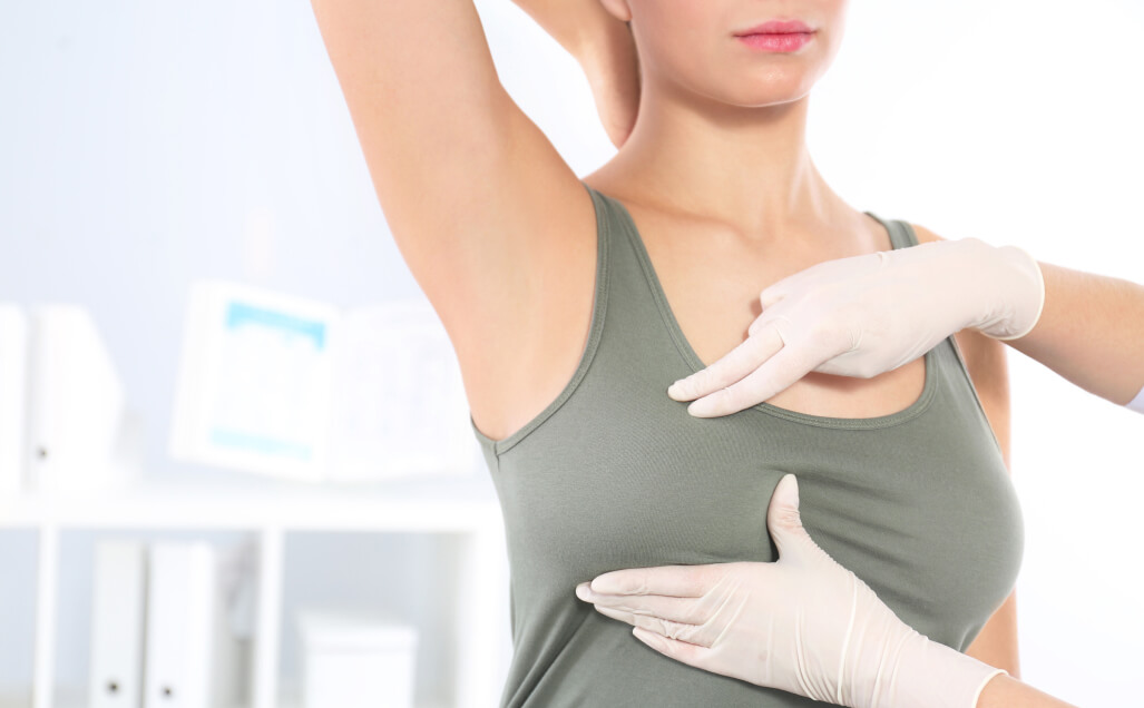
If you notice a lump in your breast or armpit area, a mammogram and ultrasound are one of the first steps in figuring out the cause of the lump. We perform ultrasounds here in the office during your visit, and will also refer you to the appropriate imaging center for mammogram and any other appropriate imaging study.
For patients with large, painful and/or bothersome cysts (benign fluid-filled sacs), ultrasound-guided cyst aspirations can be performed in the office. This is done under local anesthesia (numbing medicine). Under sterile conditions, a needle will be passed into the cyst under ultrasound visualization and the fluid pulled out into a syringe. Sometimes the fluid will be sent to the lab for analysis.
For any mass or lump in the breast or axilla that is visible on ultrasound, we can perform ultrasound-guided core needle biopsies or fine needle aspirations in the office. These procedures are done under sterile conditions with local anesthesia (numbing medicine) and you are able to drive afterwards. Specimens are sent to the pathology lab for analysis. The procedure takes approximately 1 hour.
Small benign masses, such as fibroadenomas, can sometimes be removed without any open surgery. This can be done in the office using the core needle biopsy device. This biopsy device removes cores of tissue and thus can effectively remove an entire benign mass through a small incision. The procedure is done under local anesthesia (numbing medicine), and you will be able to drive afterwards. The procedure takes approximately 1 hour
For patients who have a long-term tunneled venous access device (port/portacath/mediport), we can remove most of these in the office under local anesthesia. If the patient desires sedation (twilight sleep) or general anesthesia during the port removal, the procedure will be done at the hospital. When done in the office, the procedure takes approximately 1 hour.
Surgical Procedures
Benign masses or non-cancerous abnormalities (atypia) are removed surgically in the operating room. When the area to be removed is not easily felt, a wire or metallic seed is placed in the area to guide the surgeon to the area that needs to be removed. The wire or seed is placed in radiology under local anesthesia. The excisional biopsy is an outpatient surgery and done usually under general anesthesia.
Lumpectomy is performed as part of treatment for breast cancer. This involves removal of the cancer with a rim of normal tissue (clear margin). If the cancer cannot be easily felt, a wire or metallic seed is placed prior to surgery into the area of cancer to the guide the surgeon (done in radiology with local anesthesia). This is an outpatient surgery and usually done under general anesthesia.A lumpectomy can often be performed as a Hidden Scar procedure. This means that the incision is made in a place that is hard to see. There are three different locations for a lumpectomy incision that make the scar less visible:
- Inframammary Fold – The natural crease beneath the breast
- Periareolar – Along the edges of the areola
- Axilla – In the armpit, usually hidden in a natural fold
Oncoplastic surgery combines sound oncologic principles with plastic surgery techniques to provide excellent cosmetic results without compromising outcome. The priority will always be providing the best cancer outcome/survival, but we aim to do so with the best aesthetic results. We offer lumpectomy with hidden scars and oncoplastic closures to minimize dimpling and retraction of the skin. We work with our plastic surgery colleagues to provide reconstructive options such as simultaneous breast reductions at the time of lumpectomy, implant based reconstruction and autologous tissue reconstruction (using your own tissue) from the abdomen, back, or thigh.
- Oncoplastic flap advancement closures are performed during lumpectomy to bring the surrounding breast tissue together to minimize the dimpling and concavity of the breast where the cancer was removed. A Biozorb marker is also sutured into the cavity to fill the space and mark the lumpectomy cavity for radiation treatment.
- Hidden scar techniques use an advanced approach to removing breast cancer. We pride ourselves on providing our patients with the most advanced surgical options in breast cancer surgery. Our surgeons have undergone extensive training in advanced surgical techniques, and together, our goal is to give you the best surgical treatment and experience. Want more information on Hidden Scar Breast Cancer Surgery? Visit breastcancersurgery.com to learn more.
Mastectomy is performed for the treatment of breast cancer and also for risk reduction for women who carry a genetic mutation. Mastectomy involves removal of the entire breast. There are several mastectomy options: a simple (total) mastectomy, a skin sparing mastectomy, and a nipple sparing mastectomy (NSM). For patients who are not interested in reconstruction after mastectomy, excess skin is removed with the breast tissue to provide a smooth chest wall so that a breast prosthesis can be easily worn after healing is complete.
- A Nipple Sparing Mastectomy can often be performed as a Hidden Scar Procedure. This means that the incision is made in a place that is hard to see. Nipple and skin sparing mastectomy leaves the nipple areola complex and the skin envelope intact to provide the best cosmetic outcome. This is combined with reconstructive surgery by our plastic surgery colleagues, either by implant or using your own tissue (autologous tissue reconstruction). This procedure provides an excellent cosmetic outcome. However, the skin and nipple will usually be numb.
For patients with invasive breast cancer, a lymph node biopsy is generally recommended at the time of either lumpectomy or mastectomy. This is done to be sure the cancer in the breast has not spread to the lymph nodes under the arm. The word sentinel means first, but it does not always mean one. Every person has a different drainage pattern from the breast to the lymph nodes. Sometimes the breast drains only to one node before going to the rest, but other people have a few lymph nodes that drain the breast first before going to the rest. Your surgeon will not know how many sentinel lymph nodes you have until the time of surgery. Before surgery, you will have a dye injected into your breast that will drain through your breast and get stuck on your sentinel node(s). Your surgeon will identify that node(s) and remove them at the time of surgery, leaving the rest of your nodes in place. The risk of lymphedema with this procedure is very low. If patients are found to have multiple lymph nodes positive, sometimes it is recommended to remove the rest of the lymph nodes, called an axillary lymph node dissection.
For some women with breast cancer who plan to undergo lumpectomy/partial mastectomy, intraoperative radiation therapy may be an option. This allows the surgeon and radiation oncologist to provide a dose of radiation to the lumpectomy site at the time of surgery. For some women, this may be the only radiation they will need. For other women, they will need additional radiation to their breast after surgery to further reduce their risk of recurrence, but may need only a shorter course of post-surgery radiation therapy. We use the Zeiss Intrabeam, and our physicians have been part of trials that have shown the efficacy of intraoperative radiation.
Ports, portacaths, or mediports are devices that are placed under the skin of the chest, just underneath the collarbone, to provide access to the vein for medication infusions and blood draws. The port that sits underneath the skin is attached to a catheter or tube that is tunneled into one of the larger veins in the body (jugular vein or subclavian vein). Patients with a port can avoid having needles or IV’s placed in their arms each time they need infusions or blood draws. The port is placed in the operating room while you are under sedation (twilight sleep) and is an outpatient surgery.
Breast Reconstruction
Patients should know that they have options when it comes to which type of surgery to have to treat their breast cancer. We encourage patient education to help guide you to your most optimal personal choice. We work closely with excellent reconstructive plastic surgeons who can offer the following procedures. When desired or necessary we can refer you to have discussions about all options, so you feel you are choosing the reconstructive option that is best suited for you.
A breast reduction or breast lift, also known as a mastopexy, is a procedure that removes part of the skin and as much breast tissue as needed to lift the nipple back up to the desired location and give good breast contour and shape. For a woman with breast cancer who has large sagging breasts, often it is an option to have your breast surgeon perform a lumpectomy (remove the cancer in the breast with a rim of normal tissue) and at the same time have your plastic surgeon lift and reduce the breast while also performing a breast reduction on the other breast for symmetry. For patients where radiation is recommended, you can proceed with radiation to the breast after having a reduction.
For patients who will undergo a mastectomy on one side only, sometimes the plastic surgeon will recommend or offer a breast lift or reduction to the other breast to help with symmetry.
Implant reconstruction after mastectomy is generally done as a two staged procedure. At the time of the mastectomy, after the breast surgeon has removed your breast, the plastic surgeon will come in and place a tissue expander behind your chest wall muscle. This is done to create a pocket where the eventual implant will sit. It is used to help allow your skin to stretch and grow to the size you desire to be with your true implant. Every week or so your plastic surgeon will put saline into the expander. Once it is expanded to the size you desire, you will be taken back to the operating room for an outpatient procedure to have the expander removed and the permanent implant placed.
Implants can be saline or silicone. It will be up to you to discuss the risks and benefits of both options with your plastic surgeon to help you decide what you want.
Some women who may desire autologous flap reconstruction but will need radiation may have a tissue expander placed at the time of mastectomy. This will allow the skin to stretch and grow prior to radiation. The radiation will then be delivered to the chest wall while the expander is in place. Once radiation has been delivered, and once your plastic surgeon feels you are healed enough after radiation, you can return to the operating room where the expander will be removed and you will undergo autologous flap reconstruction.
Autologous flap reconstruction means using your own body tissue to create a new breast after a mastectomy. These flap can be created from multiple areas of your body, with the most common being your belly fat or your back fat. Depending on the type of flap, sometimes only fat and skin make up the flap, and sometimes the flap contains fat, skin and muscle. The main advantage of having a flap is that they often feel and may look more natural than implant reconstruction. Patients will essentially undergo a tummy tuck if they have their belly fat moved to create the breast. However, this is a longer operation than having a tissue expander or implant placed, and it requires a longer hospital stay with generally 1-2 nights in the ICU for close monitoring of the flap.
Flap reconstruction is not always a good option for all patients undergoing mastectomy, so it is important to discuss this as an option with your plastic surgeon to see if they think you are a good candidate.
For women undergoing a mastectomy who may not be ready to decide upon breast reconstruction, you can still have reconstruction later on down the road if desired. Sometimes there are reasons to wait a certain amount of time after treatment (especially if you have had radiation to your chest wall), however the plastic surgeon will evaluate you, your treatment that you have had, and your healing to determine the best timing for you to have reconstruction.
Fat grafting is a procedure that can be done to help improve the aesthetic appearance of a dent, dimple, wrinkle, or asymmetry that may have occurred after a lumpectomy or mastectomy. It is designed to add volume to areas that have lost volume by using your own fat from another area of your body and moving it to the desired area in your breast. Sometimes this may require the procedure to be done multiple times for the most desirable result. One downside to fat grafting is that you can form fat necrosis, which can feel like a lump. Generally this can be viewed on ultrasound and followed. Rarely does this cause concern for cancer recurrence, but in this case a biopsy may be required.
Schedule Your Appointment at Epic Care Today!
If you are looking for comprehensive and skilled breast health care, contact Epic Care with locations in Antioch, Dublin, Emeryville and Walnut Creek, CA. Our experienced staff will ensure you receive the care and breast cancer surgery you need and deserve. To find a doctor near you, in a particular sub-specialty, please use our find your doctor feature. We hope to hear from you soon and begin your journey to restored health.